Plant problems · leaf scorch
Diagnose Hydrangea Leaf Scorch
Diagnose hydrangea leaf scorch by comparing brown margins, daily wilt, frost timing, leaf spots, and powdery growth before changing care.
Published Last updated
In this guide
- Read the pattern before treating the plant
- Use pattern, timing and moisture to separate the routes
- Map where the injury began
- Separate afternoon wilt from dead tissue
- Check moisture below the mulch
- Use the weather record as a timestamp
- Route spots and powdery growth elsewhere
- Change only what the evidence supports
- Diagnosis
Read the pattern before treating the plant
Hydrangea leaf scorch usually appears as dry brown margins or tips, with exposed foliage often showing the first or strongest injury. Confirm the route by pairing that edge pattern with recent heat, sun or wind and moisture below the soil surface. Morning recovery, frost-timed damage, bounded spots or powdery growth sends the diagnosis in another direction.
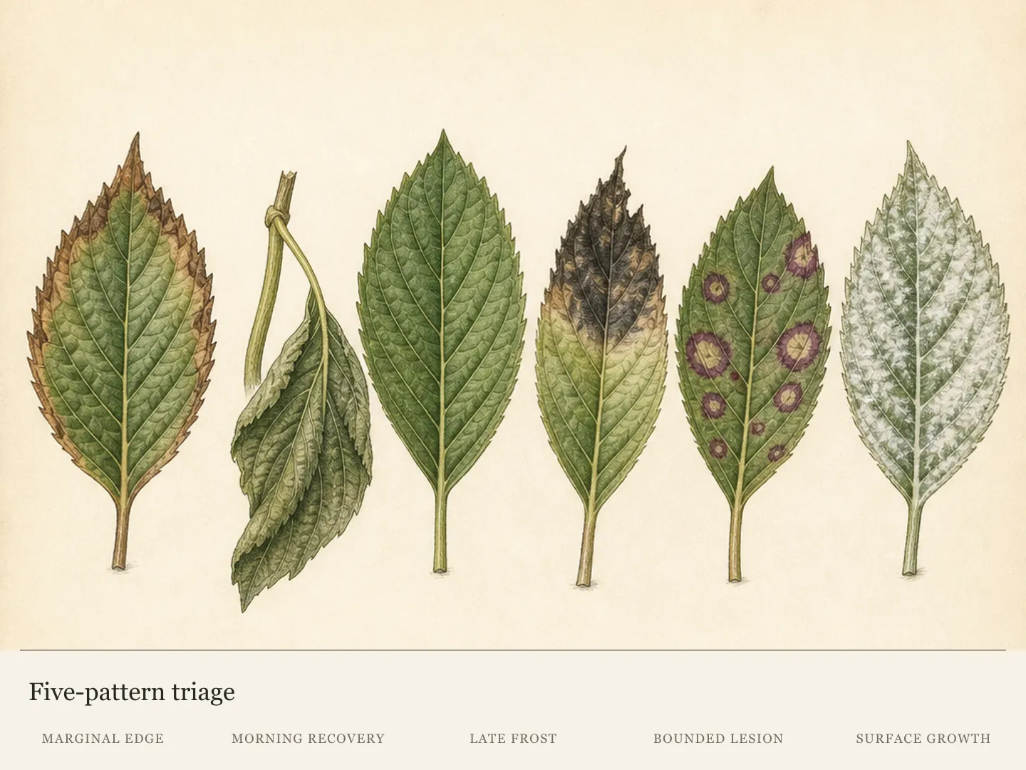
Begin with leaves still attached across the shrub. A single damaged leaf can show what tissue looks like, but it cannot reveal distribution or sequence. Compare at least ten leaves from the exposed face, shaded interior and lower canopy. Keep healthy leaves in the survey because their location may explain why one part of the plant escaped.
Brown color is the entry clue, not the diagnosis or a severity scale. A narrow crisp edge on many exposed leaves can tell a more coherent environmental story than one nearly brown leaf hidden inside the canopy. Give distribution, tissue texture and timing more weight than the most dramatic specimen, while retaining that specimen if a diagnostic service may need it.
Mark one affected shoot and photograph it late in the day and again the next morning. Record when the browning first appeared, the most recent very hot or cold night, rain or irrigation and any disturbance near the roots. This small field record preserves clues that disappear once damaged leaves are removed or weather changes.
Triage the whole plant before concentrating on leaf color. Fast spread, persistent whole-plant wilt, cane dieback or several conflicting symptom types exceeds a simple scorch check. Preserve fresh affected material and seek local Extension or plant-clinic help in that situation. Pesticides chosen before the pattern is identified can obscure evidence without correcting an environmental cause.
Use pattern, timing and moisture to separate the routes
Continuous dry margins on exposed leaves, temporary afternoon droop, injury after a cold night, bounded spots and a powdery coating lead to different checks. Pair the visible pattern with the occupied soil's moisture and recent weather before intervening. Follow new injury and next-morning posture afterward; unchanged old brown tissue does not by itself show that the correction failed.
Map where the injury began
A continuous dry edge concentrated on upper or outward-facing leaves supports environmental scorch. The inference becomes stronger when the worst foliage faces afternoon sun, reflected heat or drying wind while protected leaves remain greener. Note the compass side and height of the first damage; describing the entire shrub as simply brown discards that pattern.
Nearby pavement, a dark wall or foliage newly exposed by changes around the shrub can intensify the affected side. These site clues explain why the plant may fare worse than another hydrangea a short distance away. They also predict where new marginal injury would appear if the same exposure continues, giving the diagnosis something testable.
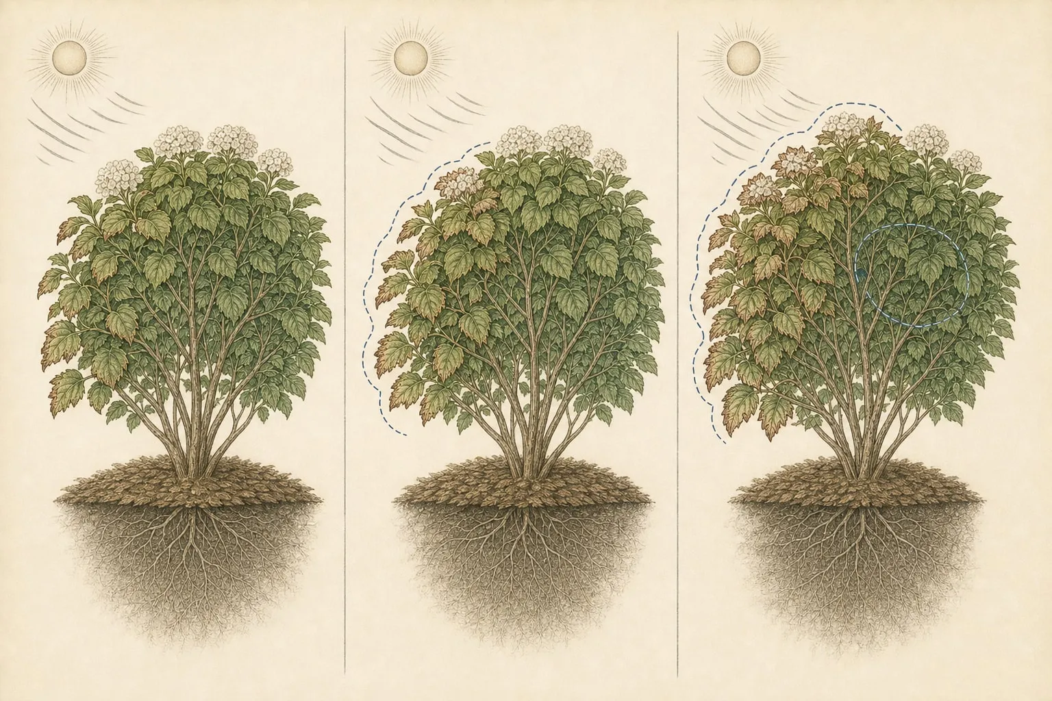
Trace the gradient from the harshest edge of the site into the protected canopy. Environmental injury may weaken across that path as reflected heat, sun or wind exposure falls. An even scatter of self-contained lesions has no comparable exposure gradient and weakens the scorch hypothesis. Record exceptions as carefully as the leaves that fit the first impression.
Scorch is a water-balance failure, not proof that irrigation was forgotten. Hot wind and sun can remove water from leaves faster than roots and vascular tissue replace it. Root limitations can produce the same imbalance even when water has recently been applied. The leaf margin records the failed supply-and-loss balance; it does not reveal which side of that balance changed.
Several pressures can operate together. A dry root zone may coincide with intense reflected heat, or a recently established root system may supply sheltered leaves while exposed ones scorch. The diagnosis should name the combined pattern when the evidence supports it. Searching for one exclusive cause can lead to repeated watering while the exposure remains unchanged, or to shade measures while the occupied soil continues to dry.
Identify the hydrangea species or cultivar if possible and describe its site. Heat, sun and dry-soil tolerance vary within the group, so a full-sun comparison from another hydrangea may be misleading. The useful comparison is within this shrub: exposed against sheltered foliage, new injury against old and root-zone condition against the weather sequence.
Separate afternoon wilt from dead tissue
A hydrangea may droop during the hottest hours and stand upright overnight. Morning recovery points toward temporary heat wilt, especially when the leaves remain green and soil below the surface is moist. It still records a demanding exposure, but it does not establish that another watering is needed. Compare the same marked shoot so normal differences between stems do not distort the result.
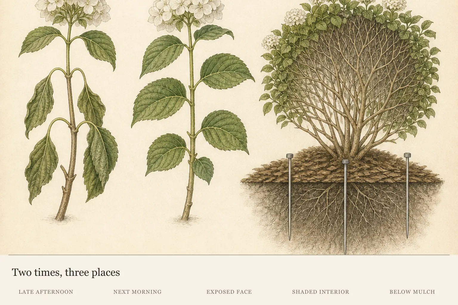
Recovery by morning is feedback, not a clean bill of health. Repeated daily wilt can still identify a site where heat load is high, and established brown edges show that some tissue has already crossed from temporary stress to injury. Use the recovered posture to prevent reflex watering while continuing to watch exposure, soil condition and the pace of new damage.
Persistent morning wilt changes the next question. Probe the root zone again and inspect the full shrub for distribution, stem condition and conflicting symptoms. Dry soil can support a watering response; cool moist soil means repeated irrigation is unlikely to resolve the heat load and may direct attention toward root function or a different diagnosis.
Crisp brown tissue is dead and will not become green again. Measure recovery through live foliage, buds and later growth while treating the old edge as a fixed historical mark. A plant can stabilize even though every scorched leaf still looks damaged. Conversely, expanding brown margins on leaves that were previously sound show that the stress process is continuing.
Appearance-based cleanup can erase that distinction. Leave enough marked foliage to compare old injury with new, and postpone hard pruning during extreme heat. The goal is to retain evidence and functioning leaf area while the plant declares whether the injury has stopped. Detached tissue can be removed without treating every marked leaf as a failing branch.
Check moisture below the mulch
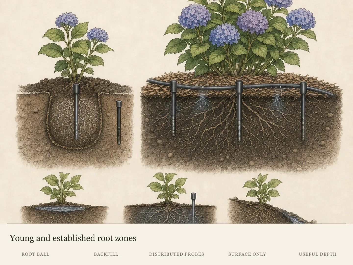
Probe at several points under the canopy without using the dry surface as a verdict. A trowel, narrow soil probe or long screwdriver can reveal whether recent water entered the root zone or merely darkened mulch and topsoil. Compare the exposed side with a sheltered point because runoff, slope and delivery can leave one zone drier than another.
A recently planted shrub requires separate checks in the original root ball and adjacent soil. Those materials can hold moisture differently while roots remain concentrated near the planting area. An established shrub calls for a broader set of points across the occupied root zone. Mixing those two stages into one test can miss the soil that is actually supplying the plant.
One moisture reading is a snapshot. Return to the same points after watering and as heat continues, noting where the probe meets cool moisture and where the profile dries first. This repeat map shows whether delivery reached the intended soil and whether one exposed side loses moisture sooner. Moving the test location each time destroys that comparison.
Dry root-zone soil paired with marginal injury supports a slow application across the useful root area. Watch whether water infiltrates or runs away, then reopen a test point after the application. A wet surface is not the checkpoint; verified moisture below it is. Repeat delivery only when the follow-up soil evidence shows that the intended zone remains dry.
Cool moist soil calls for a pause. Flooding can leave saturated or injured roots unable to meet leaf demand, so more water is not a universal answer to wilt or brown edges. Preserve organic mulch without banking it against stems. During exceptional heat, temporary overhead shade may reduce acute exposure when it stays clear of foliage and permits air movement.
Use the weather record as a timestamp
Late spring frost targets soft, newly expanded hydrangea leaves. Injury may look blackened, tan, burnt or crisp along the edges, so color alone overlaps summer scorch. The useful evidence is sequence: symptoms appeared directly after a cold night, tender tips received the injury and older or sheltered leaves escaped more of it.
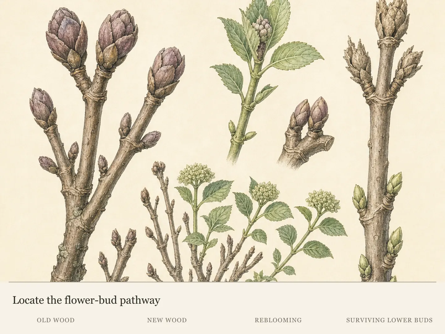
Frost injury is fixed at the tissue that was exposed during that event. Wait for live growth to show its boundaries before pruning. If later leaves emerge normally while the original tender growth remains marked, the timestamp still fits. New marginal damage during warm weather would require a fresh explanation; the old frost label no longer covers it.
Heat injury follows another sequence. Margins or exposed patches dry during or shortly after hot, sunny or windy weather, often with the greatest damage beside heat-storing surfaces. Pair that timing with canopy position and soil evidence. Heat in the forecast raises suspicion, but moist-depth checks and morning comparisons determine whether irrigation belongs in the response.
Existing brown tissue can no longer report the shrub's current water status. It remains after weather moderates and after root-zone conditions improve. Mark a few affected leaves at the edge of the damaged area and use surrounding live tissue as the forward-looking checkpoint. Stable boundaries support a stopped event; expansion into previously green tissue signals continuing injury.
When no cold or heat event fits, reopen the case. Check drainage, root disturbance, recent sprays and whether the marks are continuous edges or bounded lesions. A diagnosis that cannot explain when the damage began, where it is strongest and what new tissue does next is still provisional.
Route spots and powdery growth elsewhere
Bounded spots inside the blade behave differently from a continuous dry margin. Hydrangea leaf spots may be tan, gray, reddish brown, purple-edged or angular along veins. Inspect upper and lower surfaces and map whether lesions begin low in the canopy, follow splash patterns or occur independently of the sunniest edge.
Keep irrigation off spotted foliage while preserving root-zone care. Splash and prolonged leaf wetness can contribute to some hydrangea leaf-spot problems, so an overhead response chosen for presumed scorch may complicate the alternative diagnosis. Photograph both leaf surfaces and the whole shrub, collect fallen diseased leaves and retain a fresh attached sample if spotting spreads.
White or gray surface growth, especially with curled, puckered, yellow, red or purple leaves, points toward powdery mildew. Look beneath the same leaf whose upper surface has changed color; paired surfaces are more useful than unrelated close-ups. Powdery growth changes the investigation even though a field photograph cannot identify every pathogen.
Mixed evidence deserves preservation, not forced sorting. A heat-stressed hydrangea can also carry a foliar disease, and older damage may remain beside newer symptoms. Label samples by canopy position and date so a dry edge, bounded lesion and powdery coating are not presented as one anonymous brown leaf. The diagnostic question is whether one cause explains the set or several routes are active.
These routes matter because environmental scorch, leaf spots and powdery mildew do not share one remedy. A pesticide cannot repair abiotic scorch, and water added for wilt cannot identify a foliar disease. Local Extension or a plant clinic can evaluate a fresh sample when the pattern is mixed, advancing or too incomplete for a confident field classification.
Change only what the evidence supports
Match the action to the route with the strongest combined evidence. Dry root-zone soil plus exposed marginal injury supports slow, verified watering and relief from acute heat load. Green leaves that recover by morning over moist soil support observation. Frost-timed injury calls for patience while live growth establishes the true boundary of damage.
Bounded lesions or powdery growth need their own diagnostic path. Preserve samples, avoid creating extra leaf wetness around spots and seek local identification before choosing disease treatment. This keeps a visual resemblance from becoming an unsupported pesticide decision and protects the evidence a diagnostic service needs.
Make one evidence-matched change at a time when the shrub is stable enough to observe. A verified dry root zone can be watered; an acute exposed side can receive temporary shade; spotted foliage can be kept out of irrigation splash. Simultaneous watering, pruning and spraying would make the next observation impossible to interpret and could remove the tissue needed for diagnosis.
Repeat the same photographs and moisture checks over the next several days. Old brown tissue should remain brown, so use the rate of new injury, stability of live foliage and morning posture as checkpoints. Damage slowing after the relevant correction supports the working diagnosis without proving that every contributing factor has been found.
Write the working diagnosis as a prediction before the next check. Environmental scorch should remain strongest on the exposed side and slow when root-zone supply and acute heat load are addressed. Temporary wilt should recover by morning. Frost injury should stay tied to the tender tissue exposed during the cold event. Lesions or powdery growth should retain their distinct surface pattern. A result that breaks the prediction is a reason to reopen the route, not intensify its remedy.
A failed prediction is valuable. If margins continue to expand despite adequate root-zone moisture and moderated exposure, or if lesions and whole-plant wilt appear, stop escalating the original remedy. Bring the weather record, whole-plant view, paired leaf surfaces and a fresh sample to a local diagnostic service so the next decision begins with the full pattern.
Diagnosis
Crisp brown margins or tips are concentrated on upper and outward-facing leaves, with the strongest injury on the side exposed to afternoon sun, reflected heat or drying wind.
- Check
- Map at least ten attached leaves across exposed, shaded and lower positions. Confirm a continuous marginal pattern, match its strongest side to recent heat or wind, then probe beneath mulch at several root-zone points to distinguish dry soil from a wet surface or moist soil below.
- Cause
- Environmental leaf scorch: foliage lost water faster than roots and vascular tissue could replace it because of heat, intense exposure, drying wind, root limitation or a dry root zone. The exposed-side gradient strengthens this cause.
- Action
- When the occupied root zone is dry, apply water slowly across it and reopen a test point to verify useful penetration. Preserve crown-safe mulch and, during exceptional heat, use temporary overhead shade that remains clear of the foliage and open to air movement. Old brown edges will stay brown, so track new injury and escalate if adequate moisture fails to slow the pattern.
Green leaves droop during the hottest part of the day but regain their normal posture overnight, while the old foliage has little or no fixed brown tissue.
- Check
- Mark one green drooping shoot and photograph it late in the day and the next morning from the same angle. Pair its recovery or persistence with a below-surface moisture check; a second dose of water is unsupported when the shoot recovers over already moist soil.
- Cause
- Temporary heat wilt: short-term water loss during peak heat changed leaf posture, but the tissue recovered when conditions moderated and has not become established scorch. Repeated recovery still signals demanding exposure.
- Action
- Use the next-morning comparison as the checkpoint. Observe a recovered shoot, reduce acute afternoon exposure where practical and repeat the soil test before future watering. Moist soil calls for a pause; persistent morning wilt or whole-plant decline calls for broader root and diagnostic assessment with the time-paired photographs retained.
Soft newly expanded leaves turn black, tan or crisp immediately after a late cold night, while older leaves or growth in a sheltered position remains less affected.
- Check
- Match the first appearance of injury to a recorded late frost. Compare tender tips with older and sheltered foliage, then wait for new growth to show which buds and stems remain alive. New warm-weather damage means the frost explanation no longer covers the whole pattern.
- Cause
- Late-spring frost injury: tender new hydrangea tissue was damaged during a cold event, creating burnt-looking margins that overlap scorch in color but not in timing. Older sheltered foliage supplies the comparison.
- Action
- Let live growth define the injury before making pruning cuts. Protect tender foliage from another forecast frost where practical, retain viable stems and compare later growth with the original damaged tips. Seek help when dieback continues beyond the tissue exposed during the cold event or fresh warm-weather injury appears.
Tan, gray, reddish-brown or purple-edged marks form separate bounded lesions inside the leaf blade, while environmental scorch follows a continuous dry margin around its edge.
- Check
- Inspect both surfaces of upper and lower leaves for bounded lesions, purple halos, angular shapes along veins and a lower-to-upper distribution. Photograph the whole shrub, note splash or prolonged leaf wetness and retain a fresh sample when the spotting advances.
- Cause
- A bacterial or fungal leaf-spot problem: bounded lesions developed within the blade and may be associated with lower-canopy onset, splash or prolonged foliage wetness. A spreading case needs local identification.
- Action
- Deliver needed irrigation at the root zone without splashing the affected foliage. Collect fallen diseased leaves, preserve fresh attached samples and use local Extension or plant-clinic identification before selecting a disease treatment. Rapid spread, cane dieback or multiple symptom patterns increases the need for submitted material and a whole-plant photograph.
White or gray surface growth accompanies curling, puckering, yellowing, red or purple tissue, and the coating may be clearer on the underside of the affected leaf.
- Check
- Pair the top and underside of the same affected leaves. Look for repeated white or gray growth, colored tissue directly above it and curling or puckering on young leaves. If the pattern is mixed or spreading, preserve an intact sample for local diagnosis.
- Cause
- Powdery mildew: superficial fungal growth and associated leaf distortion or color change created a pale or powdery pattern that requires a separate disease assessment. Paired leaf surfaces clarify the route.
- Action
- Close the scorch route and assess powdery mildew on its own evidence. Relieve crowding without abruptly exposing a shade-adapted hydrangea to harsh sun, maintain root-zone care and obtain confirmation before disease-specific treatment when growth is distorted or the coating advances across untreated leaves.
Plants in this guide
Related reading

Sources
- Hydrangea: Identify and Manage Problems. University of Maryland Extension. Source record · Accessed 2026-08-15.
- How to Help Hydrangeas Weather a Heat Wave. Oregon State University Extension Service. Source record · Accessed 2026-08-15.
- Watering Established Trees and Shrubs. University of Minnesota Extension. Source record · Accessed 2026-08-15.
- What's Wrong With My Plant? Hydrangea Leaves Discolored White, Yellow or Pale Green. University of Minnesota Extension. Source record · Accessed 2026-08-15.
- Leaf Scorch of Ornamental Trees and Shrubs. University of Missouri Extension. Source record · Accessed 2026-08-15.